
World's first successful 3D imaging of living tissue with a rigid endoscope,
- Realized with an optical scanner using KTN crystal -
New Energy and Industrial Technology Development Organization
NTT Advanced Technology Corporation
Osaka Univeristy (National University)
The New Energy and Industrial Technology Development Organization (Kawasaki City, Kanagawa Prefecture, Japan, President: Kazuo Furukawa, hereafter referred to as “NEDO”) has, together with NTT Advanced Technology Corporation. (Head office: Kawasaki City, Kanagawa Prefecture, President & CEO: George Kimura, hereinafter referred to as NTT-AT) and Osaka University (Suita City, Osaka Prefecture, President: Shojiro Nishio, hereafter referred to as Osaka University), developed a rigid endoscope driven by a KTN optical scanner that is compact, high speed with a low power consumption.
Using this rigid endoscope, we have succeeded in 3D imaging of living tissue for the first time in the world. This technology enables minimally invasive diagnosis and treatment.
1. Overview
KTN (potassium tantalite niobate)*1 crystal is a material with a special electro-optic effect*2 whose refractive index changes with voltage, have the largest such effect among existing materials. By this effect, deflection (scanning) of light can be realized with a voltage of 1/100 of that for conventional materials, and by using this for a device (optical scanner) for deflecting laser light, it is possible to realize a drive that is of significantly higher speed, small size and low power consumption.
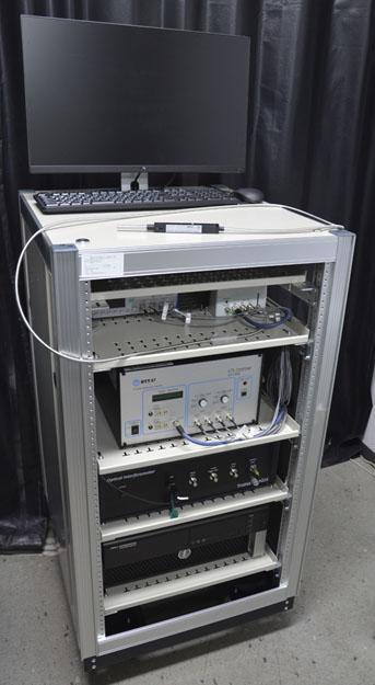
In collaboration with NTT-AT and Osaka University, NEDO, as part of its “Clean Device Promotion Project,” in partnership with NTT-AT and Osaka University, has developed a rigid endoscope*3 fitted with a newly designed lens, utilizing NTT-AT’s two dimensional optical scanner (composed of 2 KTN optical scanners). By using this rigid endoscope in combinations with Optical Coherence Tomography (OCT)*4, we have successfully succeeded in 3D imaging of living tissue using a rigid endoscope for the first time ever.
Domestically, endoscopic surgeries have already exceed 170,000 operations. This newly developed 3D imaging endoscope will be able to be deployed to the endoscopic surgery conventional up to now, as well as robotic surgery and it is expected to be used in a wide range of medical fields. In the future, NTT-AT aims to develop this device for use in a wide range of medical applications related to endoscopic surgery, including orthopedic surgery, and will proceed to provide these to medical device manufacturers as diagnostic and therapeutic devices.
2. Currently Achieved Results
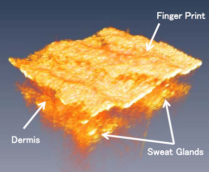
The rigid endoscope enables the acquisition of 3 dimensional images by combing the KTN optical scanner that capture the surface of the affected area in a planar manner, and the OCT that can observe living tissue in the depth direction of affected part with high definition. A lens with a diameter of 7mm and a length of 53mm is attached to the tip of the rigid endoscope to deliver laser light into the body. Imaging the affected part can be done by inserting this lens into the body from a small opening on the body surface. This time, however, we could acquire 3 dimensional images from the surface of the human finger without actually inserting the lens into the body. We were able to sufficiently image complicated internal structures such as sweat glands and dermis, so that a practical level of resolution was obtained (Fig. 2).
The rigid endoscope developed now is the result of making full use of the characteristics of [1]-[3] below of the KTN optical scanner.
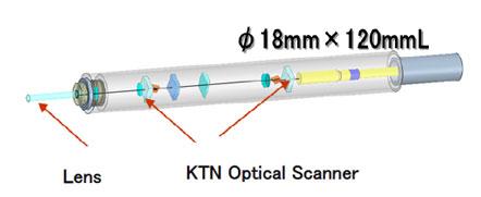
[1] Compact and Transmissive KTN Scanner
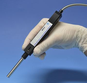
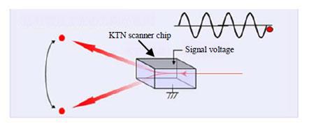
The rigid endoscope developed at this time (Fig. 5) consists of 2 KTN optical scanners (Fig. 4). In order to scan the surface of the affected area in a planar manner, a combination of the 2 KTN optical scanners is required. The typical conventional optical scanner is a reflective type that reflects laser light with a mirror such as a polygon mirror*5 or MEMS*6, whereas the KTN optical scanner is a transmission type as shown in Fig. 4, and there is no need for a complex configuration such as reflecting back the laser light. For this reason, a small number of optical parts are linearly arranged along the optical axis of the laser light to achieve low loss, even weak return light from the affected part was detected with OCT without loss and high quality imaging is possible. This simple optical system makes it possible to realize a compact, lightweight rigid endoscope of 16 mm x 183 mm in length and weighing 60g, that can withstand long-term use.
[2] Stable operation with no moving parts
The KTN optical scanner is a new type of optical scanner that controls the refractive index distribution inside the crystal depending on the voltage input, and therefore has no mechanical moving parts like a polygon mirror of MEMS. For this reason, it excels in long-term reliability, and runs stably even over long-term use. Furthermore, since the main body does not vibrate at all, precise operation during surgery is possible and stable imaging is achieved.
[3] Low power consumption through operation by electro-optic effect
Since the KTN optical scanner operates by the electro-optical effect and the KTN optical scanner’s crystal itself is a dielectric, current does not flow inside the crystal. For this reason, it is possible to reduce power consumption by 1/1000 compared to the currently most popular polygon mirror scanner and the Galvano scanner using convention OCT.
3. Plans from here on
From here on, we will conduct animal experiments and clinical trials to verify the device’s practicality as medical equipment. In addition, we aim to develop it into use for a wide range of medical fields related to endoscopic surgery, and as well, for use as diagnostic instruments we will proceed to develop therapeutic devices that make effective use of laser light.
[Glossary of Terms]
|
*1 KTN (potassium tantalate niobate) |
An optical crystal with a composition of KTa1-xNbxO3. It is transparent in a wide wavelength range and has a large electro-optical effect. |
|---|---|
| *2 Electro-optic effect | A phenomenon where the refractive index is changed by applying voltage. In the Pockels effect the refractive index is proportional to voltage and in the Kerr effect, it is proportional to the square of voltage. KTN shows Kerr effect. |
| *3 Rigid Endoscope | A type of equipment used for endoscopic surgery, also called a video scope. A CCD camera and light guide are built into the tip of a metallic tube of about 10mm in diameter, and the light guide illuminates the affected part, observing it with the CCD. During surgery, a small hole is made in the affected part and the metal cylinder is inserted in the body, and the operation is performed with observing the image of the affect part. |
|
*4 Optical Coherence Tomography (OCT) |
A technology used to observe structures in the depth direction with high resolution and high speed using light coherence. It is a tomography technology which can obtain images with non-contact/non-invasive means using laser light, without any concerns of radiation exposure. In recent years, it has been a strong focus in ophthalmology and cardiovascular internal medicine and has become widely used in the medical field. |
| *5 Polygon Mirror | A polyhedron shaped rotating mirror. It rotates at a speed of several tens of thousands times per minute, and it can scan the laser light quickly in one direction. They are widely used in laser printers, etc. |
| *6 MEMS | An abbreviation of Micro Electro Mechanical Systems which integrates mechanical element parts, sensors and actuator electronic circuits on a board. MEMS that incorporate mirrors and enable 2 dimensional optical scanning are widely used for projectors and similar uses. |

